- Home
- Journal Contents Downloads
- JRSBRT Downloads
- JRSBRT 9.3, p. 245-252
Product Description
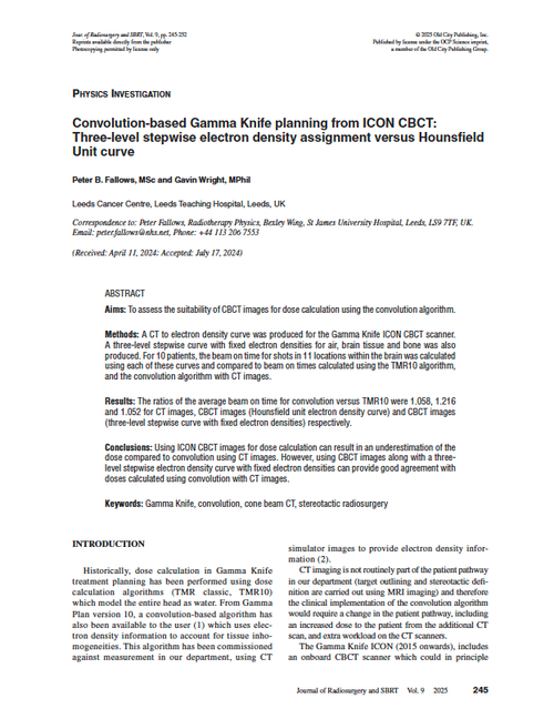
Convolution-based Gamma Knife planning from ICON CBCT: Three-level stepwise electron density assignment versus Hounsfield Unit curve
Peter B. Fallows and Gavin Wright
Aims: To assess the suitability of CBCT images for dose calculation using the convolution algorithm.
Methods: A CT to electron density curve was produced for the Gamma Knife ICON CBCT scanner. A three-level stepwise curve with fixed electron densities for air, brain tissue and bone was also produced. For 10 patients, the beam on time for shots in 11 locations within the brain was calculated using each of these curves and compared to beam on times calculated using the TMR10 algorithm, and the convolution algorithm with CT images.
Results: The ratios of the average beam on time for convolution versus TMR10 were 1.058, 1.216 and 1.052 for CT images, CBCT images (Hounsfield unit electron density curve) and CBCT images (three-level stepwise curve with fixed electron densities) respectively.
Conclusions: Using ICON CBCT images for dose calculation can result in an underestimation of the dose compared to convolution using CT images. However, using CBCT images along with a three-level stepwise electron density curve with fixed electron densities can provide good agreement with doses calculated using convolution with CT images.
Keywords: Gamma Knife, convolution, cone beam CT, stereotactic radiosurgery
After payment has been processed for your order of a digital copy (PDF) of this article, you will see a download link on your completed order page and also receive an email containing a download link. The links, which will enable you to download one copy of the article, will expire after 24 hours.
 Loading... Please wait...


