- Home
- Journal Contents Downloads
- JRSBRT Downloads
- JRSBRT 6.1, p. 11-17
Product Description
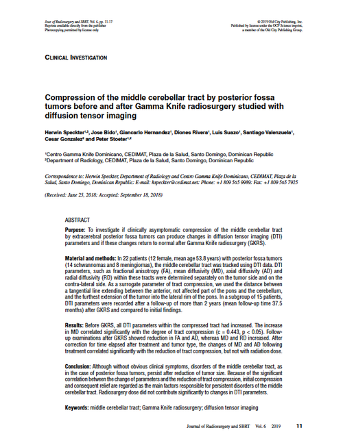
Compression of the middle cerebellar tract by posterior fossa tumors before and after Gamma knife radiosurgery studied with diffusion tensor imaging
Herwin Speckter, Jose Bido, Giancarlo Hernandez, Diones Rivera, Luis Suazo, Santiago Valenzuela, Cesar Gonzalez and Peter Stoeter
Purpose: To investigate if clinically asymptomatic compression of the middle cerebellar tract by extracerebral posterior fossa tumors can produce changes in diffusion tensor imaging (DTI) parameters and if these changes return to normal after Gamma Knife radiosurgery (GKRS).
Material and methods: In 22 patients (12 female, mean age 53.8 years) with posterior fossa tumors (14 schwannomas and 8 meningiomas), the middle cerebellar tract was tracked using DTI data. DTI parameters, such as fractional anisotropy (FA), mean diffusivity (MD), axial diffusivity (AD) and radial diffusivity (RD) within these tracts were determined separately on the tumor side and on the contra-lateral side. As a surrogate parameter of tract compression, we used the distance between a tangential line extending between the anterior, not affected part of the pons and the cerebellum, and the furthest extension of the tumor into the lateral rim of the pons. In a subgroup of 15 patients, DTI parameters were recorded after a follow-up of more than 2 years (mean follow-up time 37.5 months) after GKRS and compared to initial findings.
Results: Before GKRS, all DTI parameters within the compressed tract had increased. The increase in MD correlated significantly with the degree of tract compression (c = 0.443, p < 0.05). Followup examinations after GKRS showed reduction in FA and AD, whereas MD and RD increased. After correction for time elapsed after treatment and tumor type, the changes of MD and AD following treatment correlated significantly with the reduction of tract compression, but not with radiation dose.
Conclusion: Although without obvious clinical symptoms, disorders of the middle cerebellar tract, as in the case of posterior fossa tumors, persist after reduction of tumor size. Because of the significant correlation between the change of parameters and the reduction of tract compression, initial compression and consequent relief are regarded as the main factors responsible for persistent disorders of the middle cerebellar tract. Radiosurgery dose did not contribute significantly to changes in DTI parameters.
Keywords: middle cerebellar tract; Gamma Knife radiosurgery; diffusion tensor imaging
After payment has been processed for your order of a digital copy (PDF) of this article, you will see a download link on your completed order page and also receive an email containing a download link. The links, which will enable you to download one copy of the article, will expire after 24 hours.
 Loading... Please wait...

